Molly – 2 Year Old Labrador Diagnosed with a Torn ACL
Molly is a 2 year old female labrador who was diagnosed with a torn ACL (anterior cruciate ligament) by another veterinary clinic after playing at a park. Upon presentation at Brisbane Pet Surgery, an exam confirmed the ACL tear and testing revealed that there was a significant anterior draw sign (which is the ability to move the tibia forward comparative to the femur). The hip, hock and achilles were also assessed but were determined to be okay.
These cases are rarely x-rayed, as a clinical exam tends to be sufficient in diagnosing these sorts of injuries. X-rays won’t offer any new information, and will just cost the owner funds that could otherwise be spent on needed surgery.
A week after diagnosis, Molly was admitted to have her ACL repaired using Zlig.
What is Zlig?
Molly was admitted for LH ACL stabilisation using Zlig. She was initially placed onto IV fluids via a catheter at 290ml an hour, intubated, placed onto O2/ISO gas and given atropine via IV pre induction. Post induction, she was given betamox, metacam sc, keflin and fentanyl via IV. Post admittance, Molly had a fentanyl patch applied and she was placed onto a fentanyl infusion.
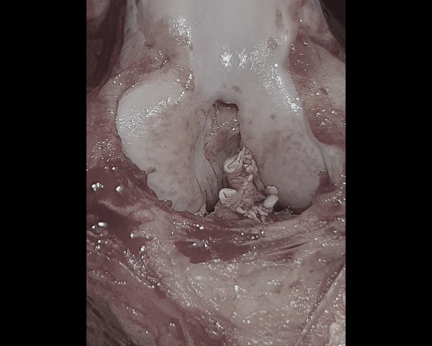
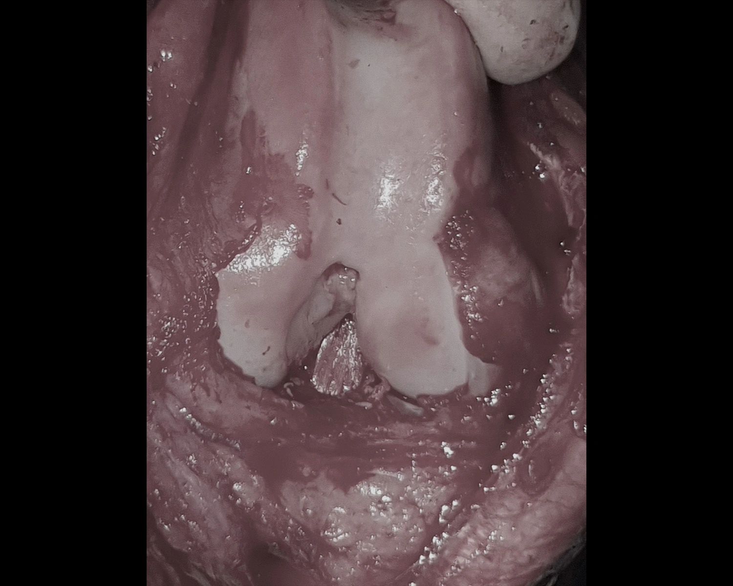
The skin over the medial LH stile was incised, a medial arthrotomy was performed and the patella was reflected laterally. The remnants of the torn ACL were identified and removed before a pin was placed through the medial wall of the lateral condyle of the distal femur. A cannulated drill bit was used to drill from the outside of the femoral condyle to the inside, at which point a pin was placed into the insertion point of the tibia and passed out further down through the tibial crest. A 25-45kg 19mm Zlig implant was passed through the tibia, through the joint and then out through the femur before a cannulated screw was placed in the femoral condyle exit.
The stifle was checked for normal movement, the implant was clamped at the exit from the tibial crest, and the anterior draw sign was checked; none were present. The cannulated screw was placed in the tibial crest, a hole was drilled in the femur around 1.5cm proximal to the condyle exit and implants were passed through this from lateral to medial. The cannulated screw was placed, the ligament end was trimmed over the medial distal femur, and a hole was drilled around 1.5cm distal to the implant exit in the tibial crest. Finally, the cannulated screw was placed and the implant was trimmed at the lateral exit from the tibia.
Procedure photos for recent case
Warning: graphic images
Molly – 2 Year Old Labrador Diagnosed with a Torn ACL
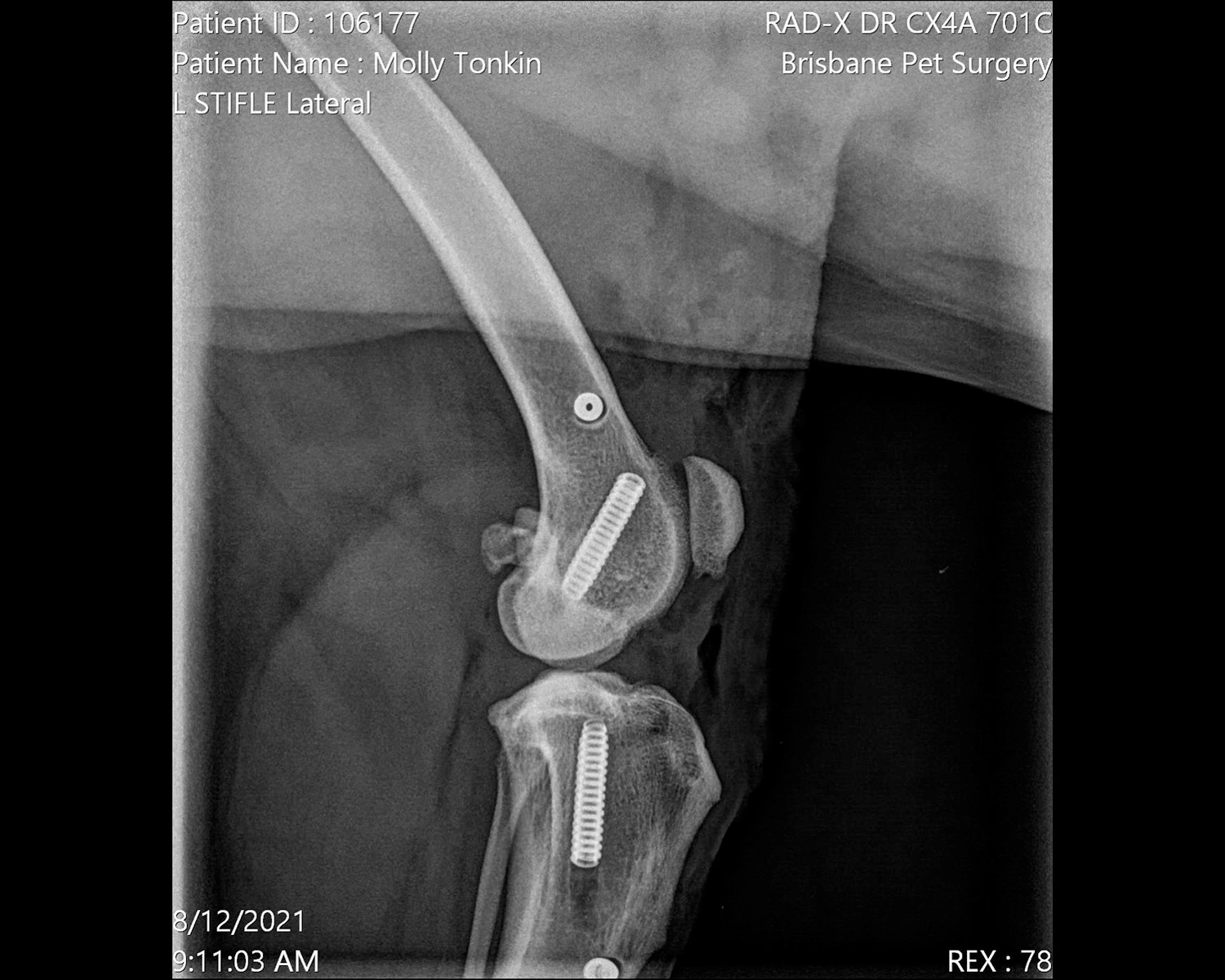


The joint was closed using absorbable sutures, then the skin was closed using absorbable ford interlocking. An x-ray of the lateral stifle was taken post op. The area was bandaged using melolin/vetwrap stik with IV fluids and fentanyl infusion continued.
Molly was able to go home later that day with strong medication to help her manage the pain (including clay tablets, gabapentin capsules, trazodone capsules, metacam oral medication and Zydax injections).
Her recovery post-surgery was fairly easy, involving rest and toilet walks only for 4 weeks. Then, for the next 4 weeks, the length of her walks was increased before she was able to go full off lead again.